Many applications in the scientific field require researchers to analyse the surface properties of materials, including their elemental, isotopic and molecular composition. Luckily, many different analysis techniques are available to scientists, and the most suitable method will depend on what they want to study. In this blog post, we will look at four of the main techniques used in surface analysis, which are secondary ion mass spectrometry (SIMS), scanning electron microscopy (SEM), transmission electron microscopy (TEM) and atomic force microscopy (AFM).
What is Secondary Ion Mass Spectrometry?
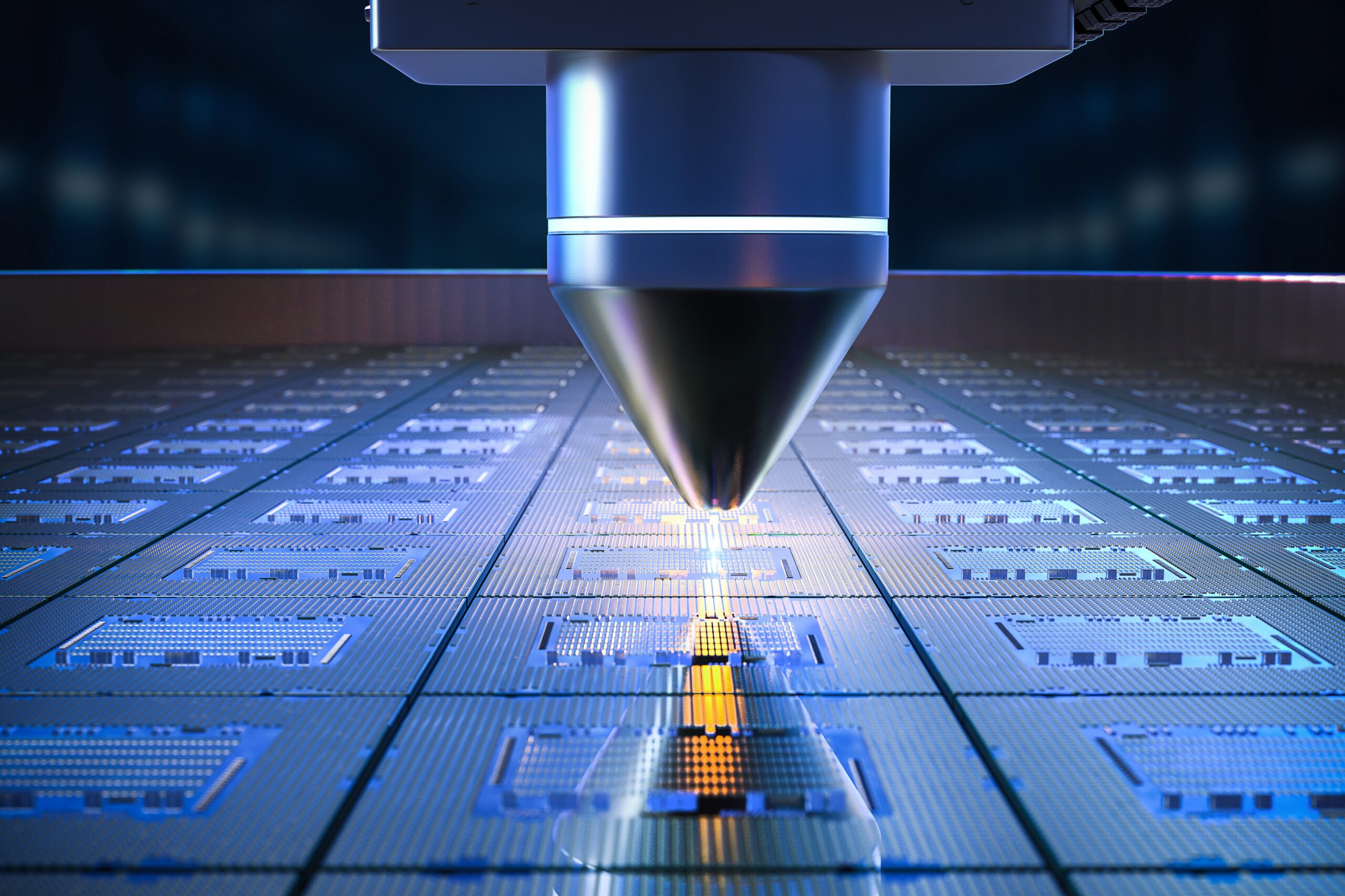
SIMS is used to analyse the composition of surfaces and thin films. The process is conducted by sputtering a material’s surface with a focused primary ion beam and then using a mass spectrometer to analyse secondary ions ejected from the sample surface. Two main SIMS analyses are available: dynamic SIMS and static SIMS, and there are three main types of spectrometers, time-of-flight (ToF-SIMS), magnetic sector and quadrupole. Which instruments are used depends on the field, such as biology, environmental science and materials science and they enable the composition of surfaces, concentration depth profiles, polymer layers and trace elements to be characterised. A valuable aspect of SIMS analysis is isotopic sensitivity enabling isotopes to be used for diffusion and tracer studies.
Scanning Electron Microscopy
Scanning electron microscopy involves scanning a sample’s surface with a focused beam of electrons, which is directed onto a small spot on the surface to create a high-resolution images. SEM produces nanoscale resolution images of nano or micro-sized materials, and enables scientists to compare tissue samples and study the composition of material surfaces. With x-ray analysis it can identify elements, but not isotopes, and the depth resolution is poorer than SIMS. X-ray detection with SEM cannot detect light elements like lithium, which are easy for SIMS.
Transmission Electron Microscopy
Transmission electron microscopy differs from SEM because it uses a wide beam of electrons, not a focused beam when imaging the sample. In addition to the beam size, TEM creates an image of a sample’s internal properties, including the morphology, composition and crystal structure. This technique is often used to analyse the cellular and molecular structures of smaller and thinner samples. It results in 2D images that, at first sight are simple but can be very rich in complex crystal structure data.
Atomic Force Microscopy
Atomic force microscopy is uses a microscopic sharp tip to detect surface topography with high levels of precision. It is a powerful technique that allows imaging and measuring many different surfaces, including biological samples, ceramics, composites, glass, and polymers and can also measure various mechanical properties of a sample. The images produced by AFM are displayed in 3D and at a higher resolution than SEM. Its other benefit is that it can analyse surface in air, not vacuum like SIMS, TEM or SEM.
Hiden Analytical and Surface Analysis
As outlined in this blog post, many techniques are available for scientists to conduct surface analysis, with the most common methods being SIMS, SEM, TEM and AFM. SIMS is used to analyse the composition of surfaces and thin films, carried out by sputtering a focused beam of primary ions at a sample’s surface and analysing the secondary ions emitted from the surface. At Hiden Analytical, we provide SIMS instrumentation for battery research, contamination, electronics, pharmaceuticals and many other applications covering multiple scientific fields.
Contact a member of the team today for more information on choosing the most appropriate surface analysis method for your applications.